Posts

Image
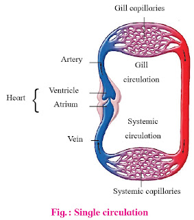
TYPES OF CLOSED CIRCULATORY SYSTEM In all vertebrates, higher molluscas and annelids, the blood is circulated all over the body through a network of blood vessels. In this type of circulation, blood flows within the blood vessels and does not come in direct contact with cells and body tissues. Exchange of material between blood and body tissues is through an intermediate fluid celled lymph. Blood flows with high pressure and contains respiratory pigments like haemoglobin for transportation of respiratory gases. Close circulation is of two types- single and double circulation. Single circulation In single circulation, the blood passes through heart only once during each cycle. ex. Fishes Deoxygenated blood is pumped from heart towards gills, where it undergoes oxygenation. This oxygenated blood moves towards various body parts, gets deoxygenated and returns back to heart for next cycle. The heart of Fish carries only deoxygenated blood, it also called 'Venous Heart'. Double Circ
Image
  MECHANISM OF ACTION OF LYMPHOCYTES
Image
The Human Heart  Introducation- The Heart is the main pumping organ of the circulatory system. It is reddish brown in colour, hollow and muscular organ. Its average weight is about 300gm in males and 250gm in female. It is conical in shape and lies in mediastenum i.e. the space between two lungs. It is broader at upper and conical at lower end (apex). Conical end is slightly tilted to left side and rests above the diaphragm. Heart is enclosed in a membranous sac called pericardium . Pericardium is formed form two main layers- outer fibrous pericardium and inner serous pericardium . Serous pericardium is soft, moist and elastic. It is formed of squamous epithelium and is further divisible into two layers as parietal and visceral layer. Parietal and visceral sublayers of serous pericardium are separated by a pericardial space. This space is filled with pericardial fluid which acts as a shock absorber and protects the heart from mechanical injuries. It also keeps the heart moist and acts

COMPOSITION OF BLOOD

Image
biologicalinform.blogspot.com

WATSON AND CRICKS MODEL OF DNA

Image
biologicalinform.blogspot.com WATSON AND CRICKS MODEL OF DNA The double stranded helical structure of DNA was proposed for the first time by James Watson and Francis Crick in 1953. In Eukaryotic, DNA molecule is present in each chromosomes if nucleus. The two strands of DNA molecule are spirally twisted around each other as well as a central axis to form tight handed double helix. The coiling of two strand is plectonemic i.e. the two strands can’t be easily separated from each other. The backbone of strand is formed by alternately arranged Deoxyribose sugar and phosphate molecular which are joined together by Phosphodiester bonds . Each strand is long polypeptide chain composed of many basic units or monomers known as Deoxyribonucleotides, hence called polymer compounds. Each transverse step is made up of nitrogen bases which are purines and pyrimidines. Nitrogen bade is attached horizontally at carbon number one of Deoxyribose sugars. The nitrogen bases of two DN

LICHENS

Image
LICHENS The lichens word was first used by Theophrastus in 300 B.C. The Lichens are the organism which form with two different partners fungi and algae. fungi and algae are in symbiotic relation which help to one another.  The lichens are distributed in wide variety of habitats. They grow on tree trunks, decaying logs, and on soil too. They are found in extreme  climatic conditions such as on dry exposed rocks and also at snow covered Arctic and Antarctic poles. They are sensitive to air pollution and generally do not grow near th cities. Their thallus are greenish or bluish green in colour, but some time yellow, orange, brown and block colour found as have additional pigments in lichens. Alexopoulos and Mims classify the lichens on the basis of fungal components like Ascolichens, Basidiolichens and Deuterolichens. On the basis of external form the lichens are of types- Leprose Lichens- The simplest type of  lichens. The thallus in which fungal hyphae envelops either si

Ultra-structure of Chloroplast

Ultra-structure  of Chloroplast (site of photosynthesis) Introduction- Schimper(1883) coined the term chloroplast for green plants. The process of photosynthesis takes place in green plastids called chloroplasts (Cholos-green & Plastos-moulded). Chloroplast are present in mesophyll cells of leaf. Shape- chloroplasts are spherical, oval or disc shaped in higher plants. The shape varies in algae. Exam- Spirogyra -spiral shaped, Zygnema -star shaped, Ulothrix -girdle. Size- chloroplast measure about 4-10 μm in length, 1-5 μm broad and 2-4 μm in thickness. Number- in some algal cells there is only single chloroplast. In higher plants the number is large. There are 20-40 chloroplast in each cells. Distribution- chloroplast are evenly distributed throughout the cytoplasm. Chemical composition- chloroplast contain 50-60% protein, 23-25% phospholipids, 5-10% chlorophyll, 2-3% RNA, 1-2% other pigments, .5% DNA, vitamins and metal ions in traces.